

Researchers Identify High-IgA MASLD Subtype Linked to Liver-Related Events
- Share
- Tweet
- Send to email
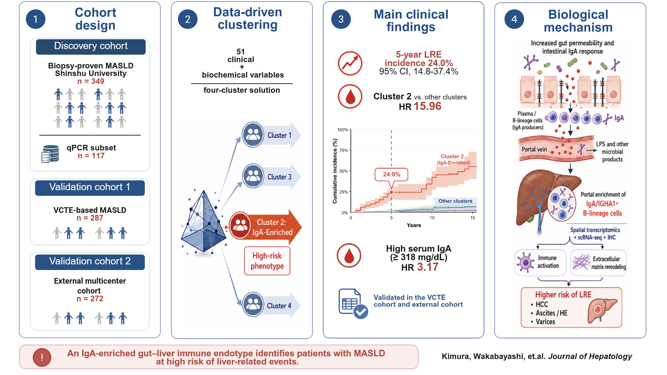
Image title: IgA-Enriched MASLD Subtype and the Gut–Liver Axis
Researchers identified an IgA-enriched subgroup of MASLD associated with increased liver-related events. Spatial transcriptomics and tissue analyses further revealed IgA-expressing B-lineage cells in portal liver regions, highlighting coordinated immune activity along the gut–liver axis.
Study reveals coordinated immune activity in the gut and liver that may help explain differences in disease progression
Metabolic dysfunction-associated steatotic liver disease can progress differently among patients. Researchers at Shinshu University identified an IgA-enriched MASLD subtype with increased risk of liver-related events, even after accounting for advanced fibrosis. The team found increased IgA-associated immune activity in the gut and liver, including IgA-expressing B-lineage cells in portal regions. The findings highlight the gut–liver axis and suggest that IgA may help identify high-risk patients for closer monitoring and treatment research.
Metabolic dysfunction-associated steatotic liver disease (MASLD) is a common liver condition that can follow very different paths between patients. Although fibrosis is a key indicator, some patients without advanced fibrosis can still develop serious liver-related events. This variation led researchers to investigate whether distinct clinical subtypes of MASLD could reveal additional clues about disease progression and risk.
A research team was led by Lecturer Takefumi Kimura, along with doctoral student Shun-ichi Wakabayashi and Professor Naoki Tanaka, from the Division of Gastroenterology, Shinshu University School of Medicine, Nagano, Japan. The team analyzed clinical and laboratory information from 349 patients with biopsy-proven MASLD and used unsupervised clustering to identify groups with different characteristics. Four subgroups emerged. The researchers then used liver tissue analysis, single-cell RNA sequencing, spatial transcriptomics, and immunohistochemistry to examine the high-risk group. The study was published online in the Journal of Hepatology on July 15, 2026.
One subgroup, Cluster 2, stood out because patients had higher blood levels of immunoglobulin A (IgA), an antibody involved in immune defense, along with older age, lower platelet counts, and more frequent diabetes. This group also had the highest prevalence of advanced fibrosis. During a median follow-up of 6.8 years, 20 liver-related events were recorded. The 5-year cumulative incidence of liver-related events in Cluster 2 was 24.0%. A blood IgA level of 318 mg/dL or higher independently predicted liver-related events even after accounting for advanced fibrosis.
These findings were also observed in additional patient groups. In a separate cohort of 287 patients without liver biopsy, those who developed liver-related events had significantly higher blood IgA levels. An independent multicenter cohort of 272 patients reproduced the IgA-enriched subgroup and its association with liver-related events. In the original cohort, patients with both elevated IgA and advanced fibrosis had a 10-year cumulative incidence of liver-related events of 48%, compared with less than 5% in the remaining patients.
“These findings show that MASLD is not a single, uniform disease and that high IgA may help identify a subgroup with a distinct pattern of disease progression,” said Dr. Kimura. “The IgA-enriched phenotype provides another way to understand risk beyond fibrosis alone.”
The team next investigated why IgA might be associated with disease progression. In patients with MASLD and advanced fibrosis, IgA-positive cells were increased in the intestinal mucosa and submucosa. Blood IgA levels were associated with EndoCAb IgG, a marker related to intestinal barrier dysfunction. In liver tissue, spatial transcriptomics showed higher IGHA1 expression in patients with advanced fibrosis, while immunohistochemistry revealed more IgA-positive cells in portal regions. Single-cell analysis showed that IGHA1 was mainly expressed by B-lineage cells, including plasma cells. Genes found alongside IGHA1 were linked to B-cell signaling and extracellular matrix organization, suggesting a connection between immune activity and liver remodeling.
“By examining both the intestine and liver, we found evidence of coordinated IgA-associated immune activity along the gut–liver axis,” said Dr. Kimura. “This provides a basis for further investigation of the biological features of the high-risk subgroup.”
Overall, the study identifies an IgA-enriched MASLD subtype associated with liver-related events beyond advanced fibrosis and highlights the complex relationship between the gut and liver. The findings could support future approaches that combine immune features with fibrosis measures to better classify MASLD. Further prospective studies and research in diverse populations will be needed to determine how these findings could inform patient monitoring and future treatment research.
Reference
Title of original paper: IgA-enriched phenotype predicts liver-related events in MASLD
Journal: Journal of Hepatology
Related SDGs